Home » Small Animal In Vivo Imaging » Ultrasound Imaging System
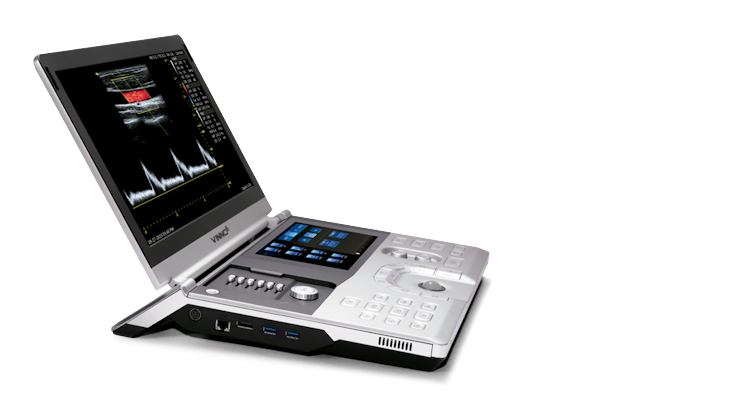
VINNO6 LAB
The world’s first portable ultrasound imaging system for preclinical research on small animals
Introduction
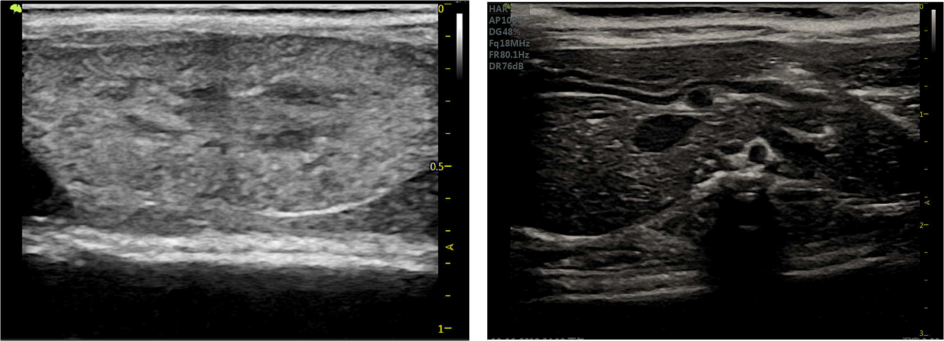
As the first portable small animal ultrasound equipment, VINNO6 LAB is a high-frequency and high-resolution ultrasound imaging system developed for experimental small animals, providing researchers with non-invasive continuous analysis, high-resolution image quality and quantitative analysis of tissue structure and blood flow velocity in foundational and preclinical studies.
VINNO6 LAB is in a compact, ultra-portable form. This lightweight system can be carried around with ease or attached to a mobile cart equipped with a multi-probe connection. Powered with Vermon high frequency probes and the innovative RF Data transmission platform, the system delivers clear images and precise measurements. It is the cutting-edge ultrasound solution for preclinical researches in biology, physiology, pharmacology, oncology and kinesiology of Cardiovascular, nervous, Developmental and Reproductive, Gastrointestinal, Pulmonary, Musculoskeletal, etc. systems, and all your other imagining needs.
Features
Portable, Durable, Low Costs The compact and ergonomic design, careful and solidly manufactured, it is very easy to carry around, setup, operate, saving money in establishment and maintenance
User Friendly, Simple To Learn User friendly interface and intuitive touch panel operation and built-in user manual tutorial, it is very simple to learn and operate, saving time and resources
Wide Range Of Application Covering a wide range of animal types, one machine can meet the application needs of all types of experimental animals, from mice, rats to dogs, pigs, etc.
Innovative RF data transmission platform The innovative RF data transmission platform collects raw data 40 times higher than traditional technology, greatly improving image contrast and resolution
Complete Functional Modules It has complete functions and can meet the needs of high-resolution imaging of the heart, blood vessels, brain, abdominal and other organs and the measurements
Precise Imaging Powered with Vermon 2-23 MHz frequency range probes it provides clear images of both superficial and deep tissues with precise measurements
Specifications
System Description:
- 15″ high resolution, high definition liquid crystal display
- 8″ touch screen operation
- 250GB (upto 2TB) SSD hard drive
- 2 USB ports
- Quick store to USB memory stick USB
- Up-to-date connectivity LAN
- Up to 1500 seconds cine storage
- Test information database
- Report package
Scanning modes:
- 2D-Mode
- B+CF
- M-Mode
- PW/HPRF Doppler
- Color Angio
- Duplex, Triplex
Full measurement and analysis package:
- Real time auto wave Doppler track and calculations
- Vascular calculations
- Cardiac calculations
- OB calculations and tables
- Gynecological calculations
- Urological calculations
- Renal calculations
- Volume calculations
Applications Cases
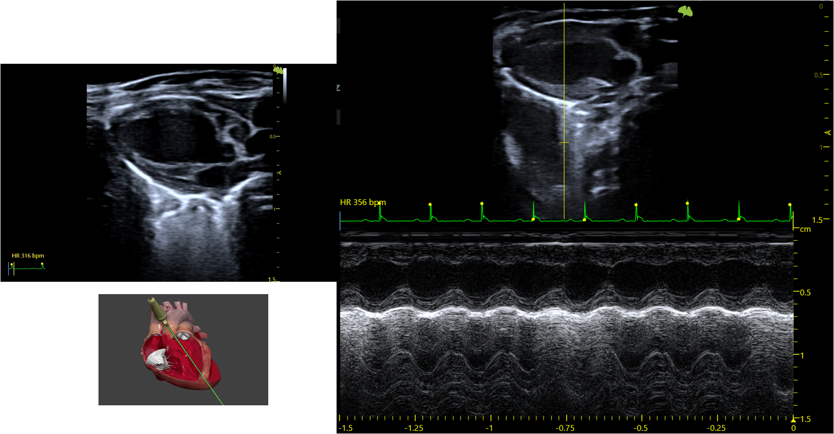
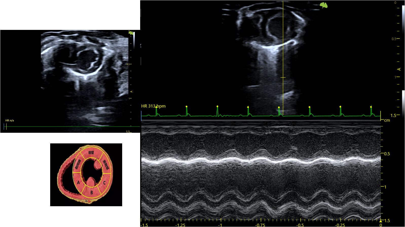
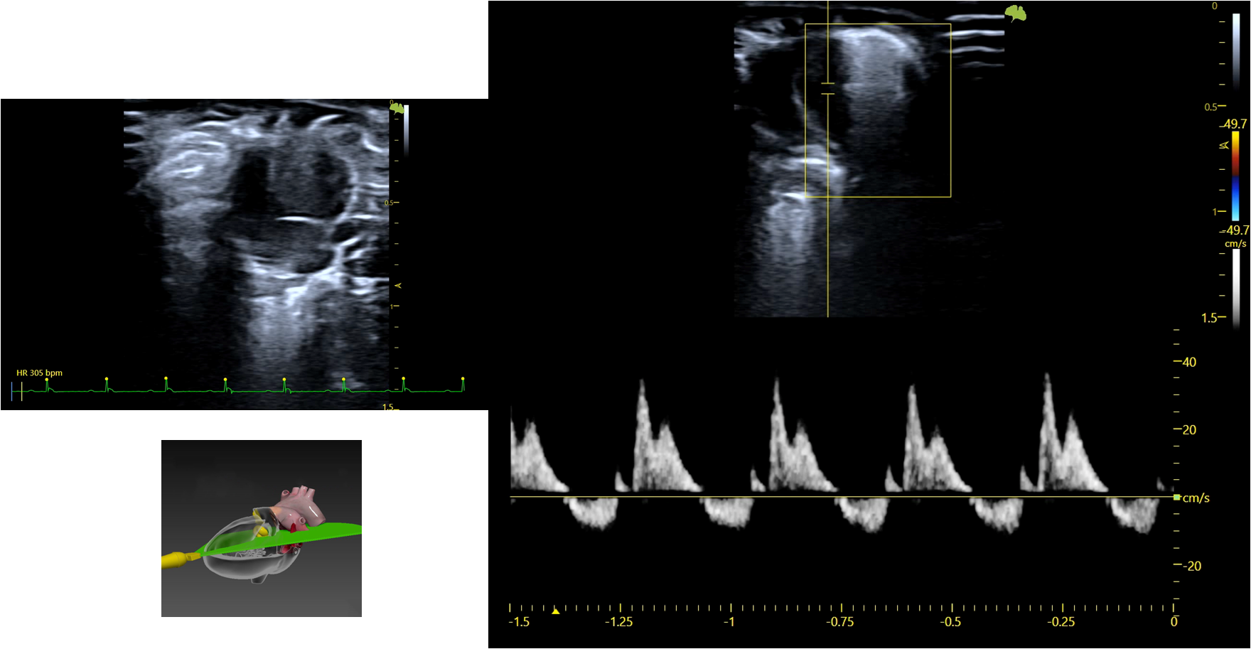
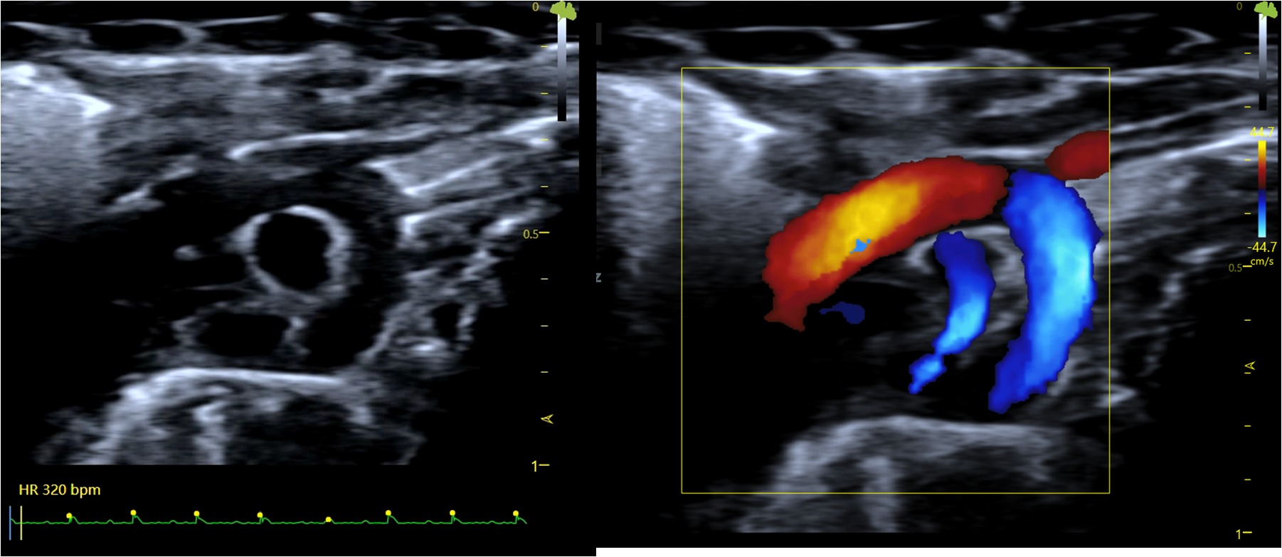
Cardiovascular
- Cardiac structure
- Congenital tissue defect
- Cardiac function evaluation
- Vascular function evaluation
Angiology
- Cross-sectional images of blood vessels
- Measurement of the thickness of the arterial wall and the size of the lumen
- Measurement of the intima, media, adventitia of the blood vessel wall
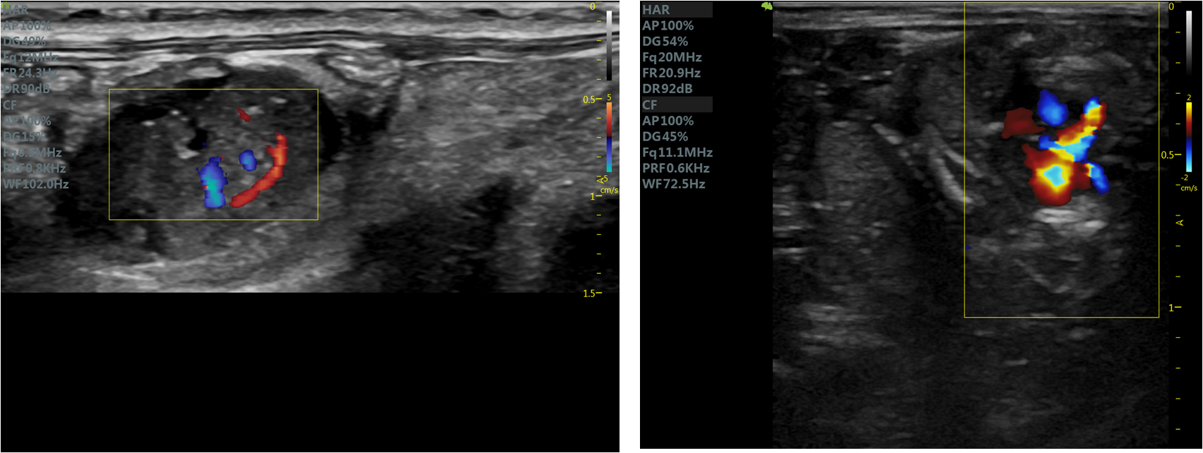
Oncology
- Tumor detection and morphology
- Measurement of parameters without the need of markers
- Oncological pharmacology
Developmental and Reproductive
- Prenatal screening
- Genetic diseases
- Reproductive function
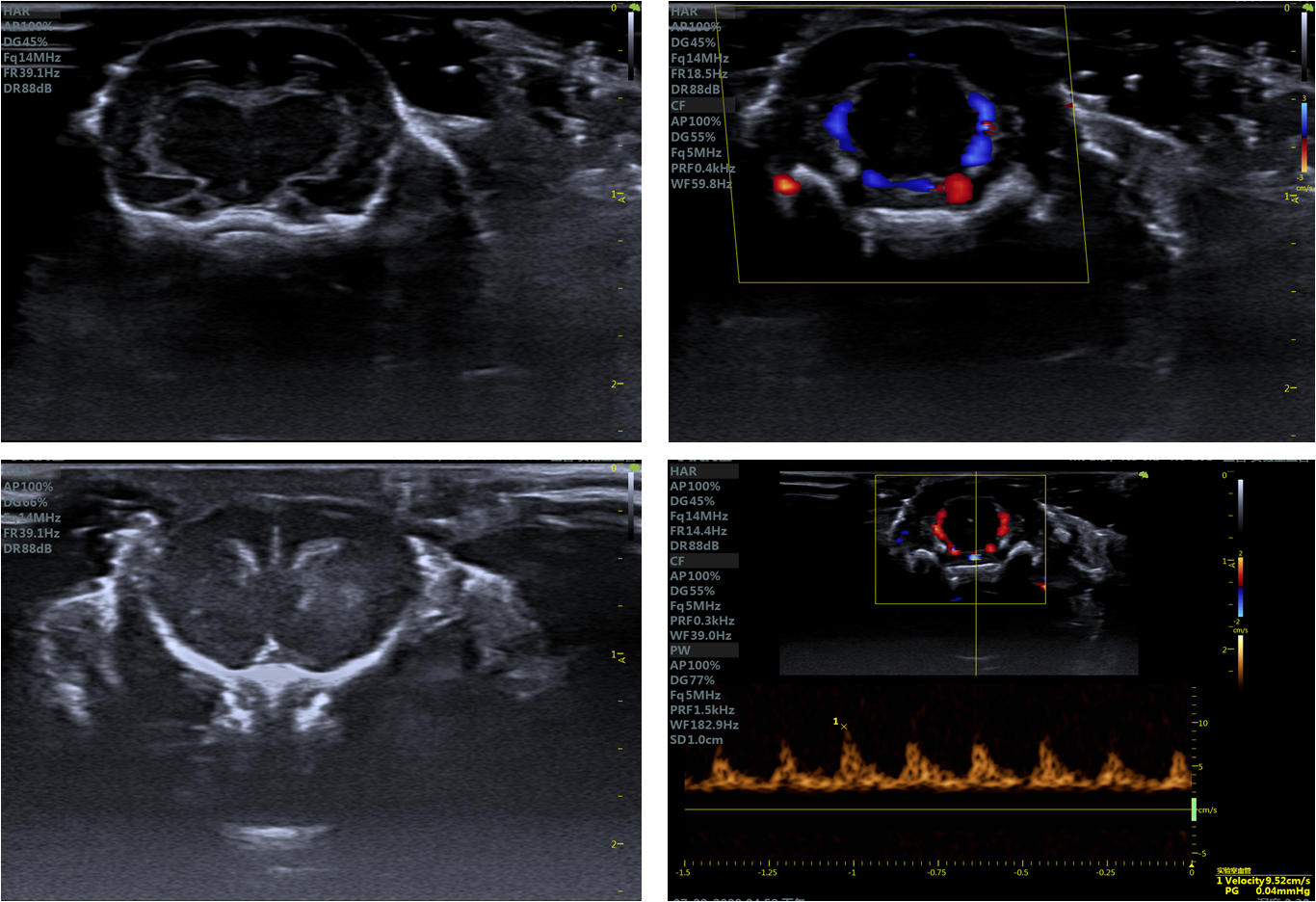
Neuroscience
- Brain tissues
- Brain blood flow
- Brain tumors
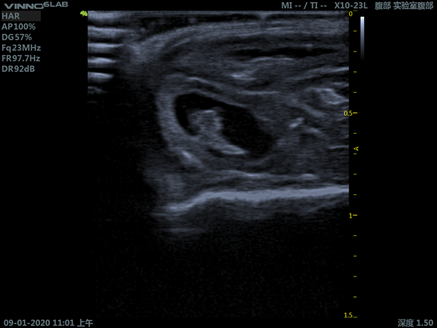
Gastrointestinal
- High-resolution two-dimensional imagings
- Real-time in vivo detection of blood flow and perfusion imaging
- Liver, kidney, intestine and other tissues and organs of various animals such as rats and mice
- Prenatal screening
- Genetic diseases
- Reproductive function
References
Since the portable model was first introduced in 2018, more than 200 sets have been in use in universities, clinical research centers, CROs and pharmaceutical R&D labs, mostly in Asian countries. Shown here are selected recent publications in peer-reviewed journals.
- α-myosin heavy chain lactylation maintains sarcomeric structure and function and alleviates the development of heart failure
Cell Res. 2023 Sep;33(9):679-698. doi: 10.1038/s41422-023-00844-w. Epub 2023 Jul 13. BAF155 promotes cardiac hypertrophy and fibrosis through inhibition of WWP2-mediated PARP1 ubiquitination
Cell Discov. 2023 May 8;9(1):46. doi: 10.1038/s41421-023-00555-x.Ultrasound-triggered microbubble destruction enhances the radiosensitivity of glioblastoma by inhibiting PGRMC1-mediated autophagy in vitro and in vivo
Mil Med Res. 2022 Feb 14;9(1):9. doi: 10.1186/s40779-022-00369-0.Treatment of infarcted heart tissue via the capture and local delivery of circulating exosomes through antibody-conjugated magnetic nanoparticles
Nat Biomed Eng. 2020 Nov;4(11):1063-1075.
doi: 10.1038/s41551-020-00637-1. Epub 2020 Nov 6.Drug-loaded balloon with built-in NIR controlled tip-separable microneedles for long-effective arteriosclerosis treatment
Bioact Mater. 2022 Dec 6:23:526-538. doi: 10.1016/j.bioactmat.2022.11.015. eCollection 2023 May.Phosphoglycerate dehydrogenase activates PKM2 to phosphorylate histone H3T11 and attenuate cellular senescence
Nat Commun. 2023 Mar 10;14(1):1323. doi: 10.1038/s41467-023-37094-8.Engineered neutrophil apoptotic bodies ameliorate myocardial infarction by promoting macrophage efferocytosis and inflammation resolution
Bioact Mater. 2021 Aug 27:9:183-197. doi: 10.1016/j.bioactmat.2021.08.008. eCollection 2022 Mar.Drug-loaded balloon with built-in NIR controlled tip-separable microneedles for long-effective arteriosclerosis treatment
Bioact Mater. 2022 Dec 6:23:526-538. doi: 10.1016/j.bioactmat.2022.11.015. eCollection 2023 May.Upconversion nanoparticles regulated drug & gas dual-effective nanoplatform for the targeting cooperated therapy of thrombus and anticoagulation
Bioact Mater. 2022 Mar 17:18:91-103. doi: 10.1016/j.bioactmat.2022.03.013. eCollection 2022 Dec.V1-Cal hydrogelation enhances its effects on ventricular remodeling reduction and cardiac function improvement post myocardial infarction
Chem Eng J. 2022 Apr 1;433(Pt 1):134450. doi: 10.1016/j.cej.2021.134450. Epub 2022 Jan 4.1-Deoxynojirimycin promotes cardiac function and rescues mitochondrial cristae in mitochondrial hypertrophic cardiomyopathy
J Clin Invest. 2023 Jul 17;133(14):e164660. doi: 10.1172/JCI164660.Alleviating experimental pulmonary hypertension via co-delivering FoxO1 stimulus and apoptosis activator to hyperproliferating pulmonary arteries
Acta Pharm Sin B. 2023 Jun;13(6):2369-2382. doi: 10.1016/j.apsb.2022.12.002. Epub 2022 Dec 8.
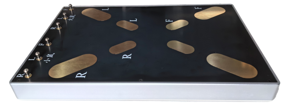
Rat and Mouse Insulated Electrocardiogram Board
It is a non-invasive ECG panel with large and uniform heating, cooperated with ultrasound imaging to monitor real-time electrocardiogram data of rats and mice and to maintain a constant body temperature during anesthesia imaging and maintain the optimal physiological state of rats and mice.
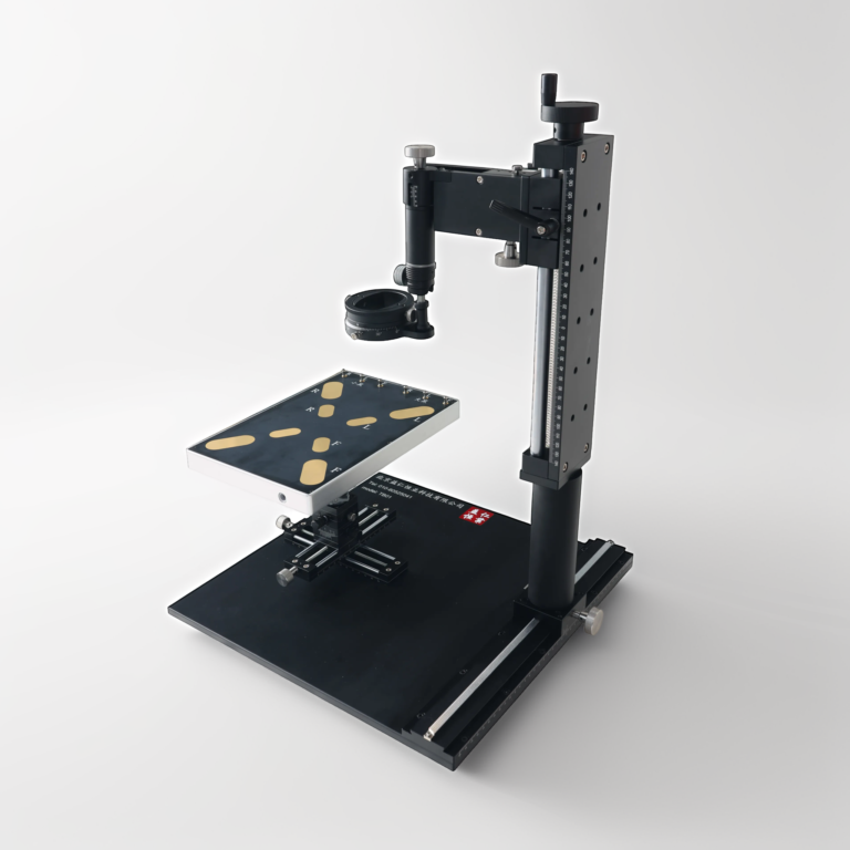
Small Animal Ultrasound Workbench
The small animal ultrasound workbench is an ultrasound imaging auxiliary tool specially customized for researchers. It can help researchers quickly set, align and adjust ultrasound probes to obtain accurate and stable ultrasound imaging. At the same time, in experiments that require long-term scanning, it can free the operator from anesthesia, achieve long-term positional stability of anesthetized animals, and allow researchers to focus more on data analysis. It is also equipped with a thermal insulation module to ensure that small animals maintain the body temperature of anesthetized animals during the anesthesia imaging process, eliminating the impact of anesthesia on the experiment, thereby improving the reliability of the experiment.
Book an On-Site Demo or Start a Free Trial
Please contact us for further information, booking an on-site demo or starting a free trial